Posts by John

Ironman Podiatrist completes Team Rothery

Ironman Wales finisher Jamie Tilley from Celtic Podiatry has recently joined Rothery Health this month from Carmarthen. Jamie will be based at Rothery Health Centre every Wednesday morning from 9-1pm. Whilst here he will be utilising his state of the art Footbalance machine to make custom orthotics (insoles) specific to you whether Ironman,footballer,runner or your weekend warrior.

What do podiatrists do ?

Podiatrists are health care professionals who have been trained to prevent, diagnose, treat and rehabilitate abnormal conditions of the feet and lower limbs. They also prevent and correct deformity, keep people mobile and active, relieve pain and treat infections.

They can give you and your family advice on how to look after your feet and what type of shoes to wear. They can also treat and alleviate day-to-day foot problems.

You can see Jamie by following the link below

http://www.rotheryhealth.com/bookings/

Becky Lewis

“After a busy year of events ending the season with two marathons four weeks apart my knees were in bits but a visit to the Magic Man John Rothery had me diagnosed with ITB Tendonitis and I was taped and treated and a treatment plan was prepared. I left his practice walking better than I had done for a month! John’s knowledge and professionalism is always top notch! Great service!”

Thanks Becky

Muscle wisdom for Tracy !


Tracy Povey who currently works at Rothery health as a therapist is currently studying for her Level 3 VTCT in Sports massage at Pembrokeshire College. She is currently enjoying learning the anatomy side of the course. Plus is revising cells,bones , body systems and postural analysis.

The content of this qualification is comprised of all the required elements needed to work effectively and efficiently as a sports massage therapist. This includes anatomy and physiology, professional practice, an understanding of the principles of health and fitness and how to provide sports massage treatments. The level 3 sports massage therapist will allow Tracy to work safely and effectively on dysfunctional tissue in a range of context, to include, pre-event, post event, intra-event and for maintenance purposes.

The structure of the qualification is comprised of 5 units which are all mandatory.
– Anatomy and physiology for sports massage
– Principles of health and fitness
– Understand the principles of soft tissue dysfunction
– Professional practice in sports massage
– Sports massage treatments

We fully support Tracy in this new venture and can’t wait for her to qualify as she will be a great adjunct to www.rotheryhealth.com


Animal Osteopathy month two!!

Animal osteopathy month two!

So this portion of the course was focused on examining and treating the hind limb. The hind limb in the horse is made up of many complex joints so there was plenty of learning to do around that as well as handling the back limb.

We also did some dissection work this month which has really opened my eyes to how complex the muscular system is and how imperative it is to keep it running smoothly in both animals and humans. 

Although we focused on hind limb, we also learnt how to examine a horse through the ‘trot-up’. You horsey people will be used to this term and what it entails, but for those who don’t, it involves trotting the horse up and down in a straight line to take note of any imbalances or any lameness. We also looked for how each joint was functioning and did it do the same things both side, and if not, why not?  

Next month we will be focusing on treating the horse through soft tissue techniques and learning about animal psychology. I will keep you posted!

In the mean-time, if you are having any aches or pains, horse riding related or not, get in touch on 07464591441 to book an appointment or look at the new online booking system at:  http://www.rotheryhealth.com/bookings

Are swimming fins dangerous?

I have noticed recently on social media sites that swimming coaches and triathletes alike have been using  terminology regarding the use of swim fins with adults and children  as “damaging” and “dangerous.”As a Health care professional and also a swimming teacher this information is misleading and therefore evidence based research is required to help inform the public. Swimming is a fantastic sport that combines all body strength, flexibility and endurance and swimmers are unfortunately prone to overuse injuries affecting the shoulder mainly, hip and knee. The ankle makes up only 1% of swimming injuries with  90% of complaints by swimmers bringing them to the doctor and/or Osteopath generally being related to shoulder problems.

Swimming with fins helps us go faster. The fin increases the surface area, as well as the kicking intensity. It makes us flex point our toes naturally, so our forward movement is faster. Consistent plantar flexion or pointing of our toes lengthens our legs muscles, especially tibialis anterior and mobilises the ankle or the talo-crural joint.Good swimmers, on the other hand, can hyperextend (plantar flex) their ankles, pointing their toes so that the top of the foot forms a straight line with the shin. Because of the extra load from the increased surface area that fins provide, swimming or kicking with fins forces ankle extension during the power phase (pushing down when swimming freestyle) of the kick. Repeated fin use eventually stretches the ankles, increasing their flexibility for moving in all directions and helping the kick become more propulsive and efficient.

The problem lies however in adults and less so in children if the ankle is restricted and fins are over used as it can lead in a few minor cases of what we call extensor tendonitis. Extensor tendinitis is inflammation of the extensor tendons which run along the top of the foot and straighten the toes. Pain is felt along the top of the foot.Treatment would involve  rest, application of cold therapy during the acute stage followed by a full rehabilitation program by a qualified osteopath or physiotherapist including gentle stretching and strengthening exercises to improve the range of motion of the ankle.

There are several factors that can predispose a swimmer to developing an injury. Your Osteopath or Physiotherapist is highly trained in identifying these and correcting them to reduce your risk of developing an injury. Some of the factors that can contribute to the development of an injury include:

  • Poor rehabilitation following a previous injury
  • Joint stiffness or swelling
  • Muscle tightness
  • Bony anomalies
  • Poor motor control and motor planning
  • Inadequate joint range of motion especially the ankle
  • Inadequate recovery periods from training and racing
  • Poor warm up / warm down
  • Over reliance on training aids
    So whilst it is important not to rely too much on training aids to improve ankle mobility there is no long term damaging or as I have heard it described as “ruining” an athlete.It is more important nonthless that coaches are more aware of abnormal kicking mechanics seen often in swimmers with knee pain rather than foot/ankle pain.For example-
  • Swimmers with medial patellar facet pain keeping their hips more abducted and utilizing greater hip and knee flexion
  • Swimmers with knee pain performing breast stroke with high angular velocities at the hip and knee and increased external tibial rotation

So in conclusion at Rothery Health we can assess you for bio mechanical weaknesses that may cause injury while swimming with fins and can treat and help improve flexibility/strength.Fins are not dangerous and can be used as a training tool . However if at any time you feel a sharp pain in your back, neck, knees or ankles whilst using them then consult one of our osteopaths immediately.

Becker, TJ (1984). The Coaches Guide to Bicepital Tendonitis. ASCA 1984 Yearbook. 71-78.

Chase KI, Caine DJ, Goodwin BJ, Whitehead JR, Romanick MA (2013). A prospective study of injury affecting competitive collegiate swimmers. Res Sports Med. 2013;21(2):111-23.

Rushall BS (2013). Relevant Training Effects in Pool Swimming: Ultra-short Race-Pace Training (Revised), Swimming Science Bulletin, 40b http://coachsci.sdsu.edu/swim/bullets/ultra40b.pdf

Rushall BS (1998). Basic Training Principles For Pre-Pubertal Swimmers, Swimming Science Bulletin, 23

Tovin BJ (2006). Prevention and Treatment of Swimmer’s Shoulder. North American Journal of Sports Physical Therapy, 1(4): 166-175.

Wanivenhaus F, Fox AJS, Chaudhury S, Rodeo SA (2012). Epidemiology of Injuries and Prevention Strategies in Competitive Swimmers. Sports Health. May 2012; 4(3): 246–251.


Want to improve performance on your horse?

My name is Lois Morgan and I have recently graduated with a Masters in Osteopathy from Swansea University and am now working with Rothery Health in Saundersfoot, Wales

I am from Swansea and have lived on a beef cattle farm my entire life and this, and my love of horse riding, sparked my interest in animal osteopathy. I am currently training to be an Equine and Canine Osteopath and hope to be fully qualified and covering the Pembrokeshire area by June 2017. The course takes me to Oxford once a month where some very generous people allow me and my colleagues to examine and practice on their horses. I have loved every second of it so far and can’t wait to be an animal Osteopath, hopefully getting these animals back fighting fit whether they are happy hackers or eventers!

Even though I have a great interest in animals, I am also treating all people in the Pembrokeshire area that are suffering with aches or pains in the. Osteopathy has the ability to help many conditions ranging from pain related to ageing and sciatica to sports injuries and headaches. I am currently offering 15 minute dropins at Rothery Health on a Tuesday for anyone wishing to discuss their conditions or any of their concerns! Call in for a chat to see if osteopathy could be beneficial for you!

Phone 07464591441 if you would like an appointment.


Lower back pain:its NOT about the chair?

As an osteopaths we often get asked what type of chair a patient with lower back pain should have .However lower back pain like most chronic painful conditions involves much more than physical factors like the way we sit, bend and lift but is reliant on other issues such a patients job satisfaction, their activity levels, their sleep, their thoughts, fears and mood .

Therefore, it is hardly surprising that changing a single thing in a persons life (e.g. the chair) without addressing any other potentially relevant factors (e.g. their job satisfaction, their activity levels, their sleep, their thoughts, fears and mood) is often ineffective.

Research has been done recently to examine the different types of chairs available on the market and it appears that while using a backrest may appear to help reduce back muscle tension to some extent, there is no clear evidence that using a backrest affects the development of Lower Back Pain. The use of a sloping chair (like a kneeler chair) did not appear to help reduce Lower Back pain either (in fact, if anything they were found to be slightly provocative).

Research also suggests that sitting on a dynamic(wobbly chair) and/or stool did not prevent lower back pain either and therefore  investing (often large sums of money) in a new chair is often NOT necessary. In summary Lower Back Pain is usually not just about the chair or indeed any other uni-dimensional factor but the factors mentioned above.

If you wish to discuss this further either call 07951381265 or visit our web site www.rotheryhealth.com

Sports Hernia or Groin strain?

Whilst on holiday in Italy recently our hotelier who was a very keen footballer/tennis player and generally a nice guy wanted to tell me all about his groin problem he had been experiencing for a number of months (once he found out I was an Osteopath….occupational hazard I suppose?) which I agreed to look at. He had been seeking various treatments from Osteopaths/GPs and Physiotherapists in Pescara,Italy and nothing appeared to be helping.

A sports hernia is probably the least understood of all the injuries that involves athletes and is a tear to the oblique abdominal muscles. Unlike a traditional hernia, the sports hernia does not create a hole in the abdominal wall and in this particular case there was no visible protrusion. As a result there is often no visible bulge under the skin and therefore this makes a definitive diagnosis of sports hernia difficult.

The soft tissues most frequently affected by sports hernias are the oblique muscles in the lower abdomen. Especially vulnerable are the tendons that attach the oblique muscles to the pubic bone. In many cases of sports hernia, the tendons that attach the thigh muscles to the pubic bone (adductors) are also stretched or torn….making diagnosis even more difficult.

A sports hernia will usually cause severe pain in the groin area at the time of the injury. The pain typically gets better with rest, but comes back when you return to sports activity, especially with twisting movements such as football/tennis. Over time a sports hernia may lead to an inguinal hernia and abdominal organs may press against the weakened soft tissues to form a visible bulge as in a traditional hernia.

On examination of this particular chap when he was asked to do a straight legged sit up there was pain in the groin area however no bulge and therefore treatment involved stretching and soft tissue treatment. He also experienced pain on coughing and on extension of the hip.Occasionally surgery to repair the torn tissues in the groin can be done as a traditional, open procedure with one long incision or as an endoscopic procedure. In an endoscopy, the surgeon makes smaller skin incisions and uses a small camera, called an endoscope to see inside the abdomen.

This indeed with rest and treatment may resolve however professional athletes often prefer to have surgery right away to resolve the problem.If you want to discuss any problem similar to this either visit our web site www.rotheryhealth.com or call

07951381265

Diagnosis of the week # Hip Impingement

As an Osteopath we often come across patients who present with unusual signs and symptoms and our job is then to assess and try to formulate a diagnosis (physiotherapists are generally given the diagnosis) .This week was no different when a lady in her mid 30s presented to Rothery Health Clinic Saundersfoot with hip pain continually which was similar to a dull ache in the region of her groin.There was no sharpness to the pain but it had progressively got worse and was beginning to bother her over a period of a month.

You can have hip impingement for years and not know it, because it is often not painful in its early stages.When hip impingement causes symptoms, it may be referred to as hip impingement syndrome. The main symptoms are stiffness in the groin or front of the thigh and/or a loss of your hip’s full range of motion which this lady was now having.

There are 3 Types of Impingement

There are three types of Impingement: pincer, cam, and combined impingement.

Pincer. This type of impingement occurs because extra bone extends out over the normal rim of the acetabulum or socket. The labrum can be crushed under the prominent rim of the acetabulum.

Cam. In cam impingement the femoral head is not round and cannot rotate smoothly inside the socket. A bump forms on the edge of the femoral head that grinds the cartilage inside causing pain.

Combined. Combined impingement just means that both the pincer and cam types are present.

With this particular lady she was starting to feel pain with more subtle activities, such as sitting for long periods of time or walking up a hills.

On examination it was clear this particular lady had reduced internal rotation of the hip and there was pain. Therefore she was referred back to her GP with a request for an MRI.

Hip Impingement occurs because the hip bones do not form normally during the childhood growing years. It is the deformity of a cam bone spur, pincer bone spur, or both, that leads to joint damage and pain. When the hip bones are shaped abnormally, there is little that can be done to prevent impingement.

Because athletically active people may work the hip joint more vigorously, they may begin to experience pain earlier than those who are less active. However, exercise does not necessarily cause impingement.

Often, surgery for hip impingement can be performed arthroscopically. This technique involves inserting a lighted scope and thin tools through small incisions over your hip instead of making a large incision.Arthroscopy is usually an outpatient surgery. This means you can go home the same day.We will now await the MRI scan however this patient was relieved in the fact that we had given her an answer to her problem and she could now move forward with her life.

If you have any issues please visit our web site www.rotheryhealth.com or call us 07951381265

7 stages to help with shoulder impingement

Shoulder impingement syndrome is a condition where your shoulders rotator cuff tendons are intermittently trapped and compressed during shoulder movements This causes injury to the shoulder tendons and bursa resulting in painful shoulder movements.

Impingement (impact on bone into the rotator cuff tendon or bursa) should not occur during normal shoulder function. When it does happen, the rotator cuff tendon becomes inflamed and swollen, a condition called rotator cuff tendonitis. Likewise if the bursa becomes inflamed, shoulder bursitis will develop.

Both these conditions can co-exist or be present independently.

While a traumatic injury can occur eg fall, it is repeated movement of your arm into the impingement zone overhead that most frequently causes the rotator cuff to contact the outer end of the shoulder blade (acromion). When this repeatedly occurs, the swollen rotator cuff is trapped and pinched under the acromion.

Injuries vary from mild tendon inflammation (tendonitis), bursitis (inflammed bursa), calcific tendonitis (bone forming within the tendon) through to partial and full thickness rotator cuff tendon tears, which may require surgery.

What Causes Shoulder Impingement?

The shoulders rotator cuff tendons are protected from simple knocks and bumps by bones (mainly the acromion) and ligaments that form a protective arch over the top of your shoulder.

In between the rotator cuff tendons and the bony arch is the subacromial bursa (a lubricating sack), which helps to protect the tendons from touching the bone and provide a smooth surface for the tendons to glide over.

However, nothing is foolproof. Any of these structures can be injured, whether they be your bones, muscles, tendons, ligaments or bursas.

Shoulder impingement has primary (structural) and secondary (posture & movement related) causes.

Primary Rotator Cuff Impingement – Structural Narrowing

Some of us are born with a smaller sub-acromial space. Conditions such as osteoarthritis can also cause the growth of sub-acromial bony spurs, which further narrows the space.

Because of this structural narrowing, you are more likely to squash, impinge and irritate the soft tissues in the sub-acromial space, which results in bursitis or shoulder tendonitis.

Secondary Rotator Cuff Impingement – Dynamic Instability

Impingement can occur if you have a dynamically unstable shoulder. This means that there is a combination of excessive joint movement, ligament laxity and muscular weakness around the shoulder joint. This impingement usually occurs over time due to repetitive overhead activity, trauma, previous injury, poor posture or inactivity.

In an unstable shoulder, the rotator cuff has to work harder, which can cause injury. An overworking rotator cuff fatigues and eventually becomes inflamed and weakens due to pain inhibition or tendon tears.

When your rotator cuff fails to work normally, it is unable to prevent the head of the humerus (upper arm) from riding up into the sub-acromial space, causing the bursa or tendons to be squashed.

Failure to properly treat this instability causes the injury to recur. Poor technique or bad training habits such as training too hard is also a common cause of overuse injuries, such as bursitis or  tendonitis.

What are the Symptoms of Shoulder Impingement?

Commonly rotator cuff impingement has the following symptoms:

  • An arc of shoulder pain approximately when your arm is at shoulder height and/or when your arm is overhead.
  • Shoulder pain that can extend from the top of the shoulder to the elbow.
  • Pain when lying on the sore shoulder.
  • Shoulder pain at rest as your condition deteriorates.
  • Muscle weakness or pain when attempting to reach or lift.
  • Pain when putting your hand behind your back or head.
  • Pain reaching for the seat-belt.

Who Suffers Shoulder Impingement?

Impingement syndrome is more likely to occur in people who engage in physical activities that require repeated overhead arm movements, such as tennis, golf, swimming, weight lifting, or throwing a ball.

Occupations that requires repeated overhead lifting or work at or above shoulder height are also at risk of rotator cuff impingement.

How is Shoulder Impingement Diagnosed?

Shoulder impingement can be diagnosed within the clinic by your physiotherapist or osteopath.

Ultrasound scan may be useful to visualise dynamic impingement and detect associated any associated injuries such as shoulder bursitis, rotator cuff tears, calcific tendonitis or shoulder tendinopathies.

During your ultrasound scan the sonographer or radiologist can visualise what is happening as your shoulder moves through the impingement zone.

What is the Shoulder Impingement Zone?

Postures that significantly narrow the subacromial space are:

  • Your arm directly overhead such as plasterers or electricians.
  • Your arm working at or near shoulder height.

shoulder impingement syndrome

Who Suffers Shoulder Impingement Syndrome?

Impingement syndrome is more likely to occur in people who engage in physical activities that require repeated overhead arm movements, such as tennis, golf, swimming, weight lifting, or throwing a ball.

Occupations that requires repeated overhead lifting or work at or above shoulder height are also at risk of rotator cuff impingement.

Shoulder Impingement Treatment

There are many structures that can be injured in rotator cuff impingement. How the impingement occurred is the most important question to answer. This is especially important if the onset was gradual since your static and dynamic posture, muscle strength, flexibility and spine shape all have important roles to play.

Once you suspect any rotator cuff injury, it is important to confirm the exact type of your rotator cuff injury since treatment does vary depending on the specific or combination of rotator cuff injuries.

Your rotator cuff is an important group of control and stability muscles that maintain “centralisation” of your shoulder joint. In other words, it keeps the shoulder ball centred over the small socket. This prevents injuries such as impingement, subluxations and dislocations.

We also know that your rotator cuff provides subtle glides and slides of the ball joint on the socket to allow full shoulder movement. Plus, your shoulder blade (scapula) has a vital role as the main dynamically stable base plate that attaches your arm to your chest wall.

Researchers have concluded that there are essentially 7 stages that need to be covered to effectively rehabilitate these injuries and prevent recurrence.

These are:

  • Early Injury: Protection, Pain Relief & Anti-inflammatory Treatment
  • Regain Full Shoulder Range of Motion
  • Restore Scapular Control and Scapulohumeral Rhythm
  • Restore Normal Neck-Scapulo-Thoracic-Shoulder Function
  • Restore Rotator Cuff Strength
  • Restore High Speed, Power, Proprioception and Agility Exercises
  • Return to Sport or Work

For more specific advice about your shoulder impingement, please contact your osteopath.

Corticosteroid Injections

Corticosteroid injections can be useful in the initial pain relieving stage if conservative methods fail to reduce the pain and inflammation. It is important to note that once you pain settles, it is important to assess your strength, flexibility, neck and thoracic spine involvement plus your scapulohumeral rhythm to ensure that your shoulder impingement does not return once your injection has worn off.

How Long Does is Your Recovery?

Every shoulder impingement is different. Some impingements will respond positively to one treatment session, whereas a more complicated case may take many weeks or a few months to settle. Others may require shoulder surgery.

There is no specific time frame for when to progress from each stage to the next. Your injury rehabilitation status will be determined by many factors during your physiotherapist’s clinical assessment.

You’ll find that in most cases, your osteopath will seamlessly progress between the rehabilitation phases as your clinical assessment and function improves.It is also important to note that each progression must be carefully monitored as attempting to progress too soon to the next level can lead to re-injury and frustration.

For more specific advice about your shoulder impingement, please contact www.rotheryhealth.com or call 07951381265

Achilles tendinitis and 5 ways to manage it

Achilles Tendinitis

Achilles tendinitis is a common condition that causes pain along the back of the leg near the heel.

The Achilles tendon is the largest tendon in the body. It connects your calf muscles to your heel bone and is used when you walk, run, and jump.

Although the Achilles tendon can withstand great stresses from running and jumping, it is also prone to tendinitis, a condition associated with overuse and degeneration.

Description

Simply defined, tendinitis is inflammation of a tendon. Inflammation is the body’s natural response to injury or disease, and often causes swelling, pain, or irritation. There are two types of Achilles tendinitis, based upon which part of the tendon is inflamed.

Noninsertional Achilles tendinitis

Noninsertional Achilles Tendinitis

In noninsertional Achilles tendinitis, fibers in the middle portion of the tendon have begun to break down with tiny tears (degenerate), swell, and thicken.

Tendinitis of the middle portion of the tendon more commonly affects younger, active people.

Insertional Achilles Tendinitis

Insertional Achilles tendinitis involves the lower portion of the heel, where the tendon attaches (inserts) to the heel bone.

In both noninsertional and insertional Achilles tendinitis, damaged tendon fibers may also calcify (harden). Bone spurs (extra bone growth) often form with insertional Achilles tendinitis.

Tendinitis that affects the insertion of the tendon can occur at any time, even in patients who are not active.

Insertional Achilles tendinitis
Cause

Achilles tendinitis is typically not related to a specific injury. The problem results from repetitive stress to the tendon. This often happens when we push our bodies to do too much, too soon, but other factors can make it more likely to develop tendinitis, including:

A bone spur that has developed where the tendon attaches to the heel bone.
  • Sudden increase in the amount or intensity of exercise activity—for example, increasing the distance you run every day by a few miles without giving your body a chance to adjust to the new distance
  • Tight calf muscles—Having tight calf muscles and suddenly starting an aggressive exercise program can put extra stress on the Achilles tendon
  • Bone spur—Extra bone growth where the Achilles tendon attaches to the heel bone can rub against the tendon and cause pain
Symptoms

Common symptoms of Achilles tendinitis include:

  • Pain and stiffness along the Achilles tendon in the morning
  • Pain along the tendon or back of the heel that worsens with activity
  • Severe pain the day after exercising
  • Thickening of the tendon
  • Bone spur (insertional tendinitis)
  • Swelling that is present all the time and gets worse throughout the day with activity

If you have experienced a sudden “pop” in the back of your calf or heel, you may have ruptured (torn) your Achilles tendon. See your doctor or osteopath immediately if you think you may have torn your tendon.

Osteopath Examination

After you describe your symptoms and discuss your concerns your osteopath will examine your foot and ankle. The Osteopath will look for these signs:

  • Swelling along the Achilles tendon or at the back of your heel
  • Thickening or enlargement of the Achilles tendon
  • Bony spurs at the lower part of the tendon at the back of your heel (insertional tendinitis)
  • The point of maximum tenderness
  • Pain in the middle of the tendon, (noninsertional tendinitis)
  • Pain at the back of your heel at the lower part of the tendon (insertional tendinitis)
  • Limited range of motion in your ankle—specifically, a decreased ability to flex your foot
Tests

Your Osteopath may request imaging tests to make sure your symptoms are caused by Achilles tendinitis.

X-rays

X-ray tests provide clear images of bones. X-rays can show whether the lower part of the Achilles tendon has calcified, or become hardened. This calcification indicates insertional Achilles tendinitis. In cases of severe noninsertional Achilles tendinitis, there can be calcification in the middle portion of the tendon, as well.

Magnetic Resonance Imaging (MRI)

Although magnetic resonance imaging (MRI) is not necessary to diagnose Achilles tendinitis, it is important for planning surgery. An MRI scan can show how severe the damage is in the tendon. If surgery is needed, your doctor will select the procedure based on the amount of tendon damage.

Treatment

Nonsurgical Treatment

In most cases, nonsurgical treatment options will provide pain relief, although it may take a few months for symptoms to completely subside. Even with early treatment, the pain may last longer than 3 months. If you have had pain for several months before seeking treatment, it may take 6 months before treatment methods take effect.

Rest. The first step in reducing pain is to decrease or even stop the activities that make the pain worse. If you regularly do high-impact exercises (such as running), switching to low-impact activities will put less stress on the Achilles tendon. Cross-training activities such as biking, elliptical exercise, and swimming are low-impact options to help you stay active.

Ice. Placing ice on the most painful area of the Achilles tendon is helpful and can be done as needed throughout the day. This can be done for up to 20 minutes and should be stopped earlier if the skin becomes numb. A foam cup filled with water and then frozen creates a simple, reusable ice pack. After the water has frozen in the cup, tear off the rim of the cup. Then rub the ice on the Achilles tendon. With repeated use, a groove that fits the Achilles tendon will appear, creating a “custom-fit” ice pack.

Non-steroidal anti-inflammatory medication. Drugs such as ibuprofen and naproxen reduce pain and swelling. They do not, however, reduce the thickening of the degenerated tendon. Using the medication for more than 1 month should be reviewed with your primary care doctor.

Exercise. The following exercise can help to strengthen the calf muscles and reduce stress on the Achilles tendon.

  • Calf stretch
    Lean forward against a wall with one knee straight and the heel on the ground. Place the other leg in front, with the knee bent. To stretch the calf muscles and the heel cord, push your hips toward the wall in a controlled fashion. Hold the position for 10 seconds and relax. Repeat this exercise 20 times for each foot. A strong pull in the calf should be felt during the stretch.

Physical Therapy. Physical therapy such as Osteopathy or Acupuncture is very helpful in treating Achilles tendinitis. It has proven to work better for noninsertional tendinitis than for insertional tendinitis.

Eccentric Strengthening Protocol. Eccentric strengthening is defined as contracting (tightening) a muscle while it is getting longer. Eccentric strengthening exercises can cause damage to the Achilles tendon if they are not done correctly. At first, they should be performed under the supervision of a Osteopath. Once mastered with a therapist, the exercises can then be done at home. These exercises may cause some discomfort, however, it should not be unbearable.

    • Bilateral heel drop
      Stand at the edge of a stair, or a raised platform that is stable, with just the front half of your foot on the stair. This position will allow your heel to move up and down without hitting the stair. Care must be taken to ensure that you are balanced correctly to prevent falling and injury. Be sure to hold onto a railing to help you balance.

Lift your heels off the ground then slowly lower your heels to the lowest point possible. Repeat this step 20 times. This exercise should be done in a slow, controlled fashion. Rapid movement can create the risk of damage to the tendon. As the pain improves, you can increase the difficulty level of the exercise by holding a small weight in each hand.

  • Single leg heel drop
    This exercise is performed similarly to the bilateral heel drop, except that all your weight is focused on one leg. This should be done only after the bilateral heel drop has been mastered.

Cortisone injections. Cortisone, a type of steroid, is a powerful anti-inflammatory medication. Cortisone injections into the Achilles tendon are rarely recommended because they can cause the tendon to rupture (tear).

Supportive shoes and orthotics. Pain from insertional Achilles tendinitis is often helped by certain shoes, as well as orthotic devices provided by your osteopath. For example, shoes that are softer at the back of the heel can reduce irritation of the tendon. In addition, heel lifts can take some strain off the tendon.

Heel lifts are also very helpful for patients with insertional tendinitis because they can move the heel away from the back of the shoe, where rubbing can occur. They also take some strain off the tendon. Like a heel lift, a silicone Achilles sleeve can reduce irritation from the back of a shoe.

If your pain is severe, your doctor may recommend a walking boot for a short time. This gives the tendon a chance to rest before any therapy is begun. Extended use of a boot is discouraged, though, because it can weaken your calf muscle.

Extracorporeal shockwave therapy (ESWT). During this procedure, high-energy shockwave impulses stimulate the healing process in damaged tendon tissue. ESWT has not shown consistent results and, therefore, is not commonly performed.

ESWT is noninvasive—it does not require a surgical incision. Because of the minimal risk involved, ESWT is sometimes tried before surgery is considered.

Surgical Treatment

Surgery should be considered to relieve Achilles tendinitis only if the pain does not improve after 6 months of nonsurgical treatment. The specific type of surgery depends on the location of the tendinitis and the amount of damage to the tendon.

Gastrocnemius recession. This is a surgical lengthening of the calf (gastrocnemius) muscles. Because tight calf muscles place increased stress on the Achilles tendon, this procedure is useful for patients who still have difficulty flexing their feet, despite consistent stretching.

In gastrocnemius recession, one of the two muscles that make up the calf is lengthened to increase the motion of the ankle. The procedure can be performed with a traditional, open incision or with a smaller incision and an endoscope—an instrument that contains a small camera. Your doctor will discuss the procedure that best meets your needs.

Complication rates for gastrocnemius recession are low, but can include nerve damage.

Gastrocnemius recession can be performed with or without débridement, which is removal of damaged tissue.

Débridement and repair (tendon has less than 50% damage). The goal of this operation is to remove the damaged part of the Achilles tendon. Once the unhealthy portion of the tendon has been removed, the remaining tendon is repaired with sutures, or stitches to complete the repair.

In insertional tendinitis, the bone spur is also removed. Repair of the tendon in these instances may require the use of metal or plastic anchors to help hold the Achilles tendon to the heel bone, where it attaches.

After débridement and repair, most patients are allowed to walk in a removable boot or cast within 2 weeks, although this period depends upon the amount of damage to the tendon.

Débridement with tendon transfer (tendon has greater than 50% damage). In cases where more than 50% of the Achilles tendon is not healthy and requires removal, the remaining portion of the tendon is not strong enough to function alone. To prevent the remaining tendon from rupturing with activity, an Achilles tendon transfer is performed. The tendon that helps the big toe point down is moved to the heel bone to add strength to the damaged tendon. Although this sounds severe, the big toe will still be able to move, and most patients will not notice a change in the way they walk or run.

Depending on the extent of damage to the tendon, some patients may not be able to return to competitive sports or running.

Recovery. Most patients have good results from surgery. The main factor in surgical recovery is the amount of damage to the tendon. The greater the amount of tendon involved, the longer the recovery period, and the less likely a patient will be able to return to sports activity.

Physical therapy is an important part of recovery. Many patients require 12 months of rehabilitation before they are pain-free.

Complications. Moderate to severe pain after surgery is noted in 20% to 30% of patients and is the most common complication. In addition, a wound infection can occur and the infection is very difficult to treat in this location.

If you require any further help please call us 07951381265 or visit our website

www.rotheryhealth.com

Ironman Wales pre and post massage special deal

Top 5 causes and solutions for ITB band pain

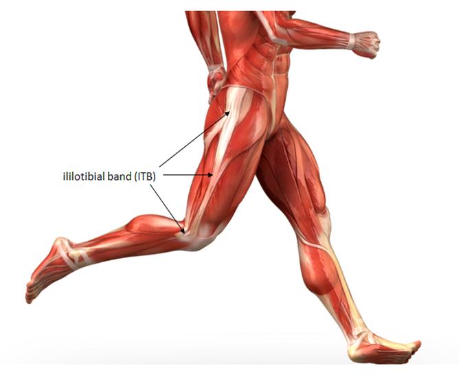
Running season has arrived and a lot of you wanted to hit the ground running, but instead, you hit the ground hurting…hurting on the outside of your knee. If that’s the case, you could have iliotibial band syndrome (ITBS) or IT band syndrome for short. IT band syndrome is an overuse injury that is common in endurance athletes like runners and bikers. It affects a tissue that runs from the side of your hip all of the way down past your knee. Most of the time, the inflammation manifests itself as pain on the outside of the knee. It can be incredibly painful and is typically a frustratingly stubborn injury to deal with. One quick look at the anatomy below can help you realize that the cause of the pain can be anywhere from the hip to the knee, so getting to the root cause can be a little tricky, but here are some solutions to a few of the most common causes of IT band syndrome.

1.  Overuse
Technically, IT band syndrome is an overuse injury. It’s kind of a misleading classification since there are usually other factors than overuse, but approaching IT band syndrome as an overuse injury is an important first step towards recovery. After icing and resting for a few days, you may need to change up your training regimen. Sometimes this can be as easy as avoiding repetition. Try changing your running route or getting away from the treadmill or track for a jog outside or go for a swim. If getting in shape or building up your endurance is your goal, try some cross-training while you slowly ramp up your mileage. Keep in mind that while overuse is usually one of the causes of IT band syndrome, there are usually other factors contributing to your pain, so some of the other interventions below may be necessary.

2.  Tight Tissues
Tight muscles in your hips or along the side of the leg can be a major contributing factor to IT band syndrome. All of these tissues are connected, so even though the location of the pain is in the knee, the hips can very well be the area that need the most attention.

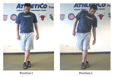
Stretching the IT band and the connected muscle is fairly simple. Just cross your legs, keeping the affected leg in back (position 1). Lean away from the painful leg until you feel a stretch along the side of the leg (position 2). Hold that for 30 seconds and repeat 5 times.
IT Band StretchIn addition to stretching foam rolling or work with a tennis ball to loosen up tight tissues and release any tightness in your hip muscles often helps. Stretch and roll both of your legs as tight tissues on one leg can cause pain on the opposite side. If there still seems to be some lingering tight spots after you try the at-home solutions, you may need to see a professional for some individualized hands-on work such as an Osteopath.

3.  Weak Hip Muscles
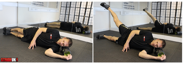
Researchers have found that weak hip muscles in particular gluteus medius can be one of the biggest reasons people get IT band syndrome. Weak muscles in the hip tend to cause your running form to break down, which puts a lot of stress on the tissues in the knee. To strengthen the muscles most commonly affected, you can try some of the exercises suggested.

side-abduction


4.  Poor Running Form
Sometimes getting rid of IT band syndrome can be as simple as changing the way that you run. Having a professional look at your running gait can reveal some problems that you may have never noticed. Some of the more common problems are overstriding (taking too long of steps) and strides that cross over the midline of the body. These are pretty easy to notice and can sometimes be pointed about by an experienced runner. Sometimes, however, the breakdown in form can be more subtle and require something more involved like a video gait analysis.

5.  Shoe or Orthotic Issues
You can correct every issue in your body, but if what comes between your foot and the ground is the cause of your problem, you’ll never see relief. Overworn shoes can cause your foot to land at awkward angles, which transfers a lot of stress up to the knee and hip, so keeping your shoes within their recommended mileage is critical. Also, adjusting to minimalist shoes (or none at all) will require you to adjust your running style, so be sure to do your research and be patient while adjusting. Finally, arch or ankle problems may require you to get orthotics so you can run with a safer gait at rotheryhealth we can assess this for you as part of the consultation.

With all of these suggestions, you can expect for improvement to take some time. If your pain lingers for longer than a few weeks, it may be time to talk to a healthcare provider such as an Osteopath for some more individualized treatment.

www.rotheryhealth.com

5 top tips to deal with post Ironman blues

This is something that most triathletes deal with, but not one that a lot of people talk about.

The cause of post-race blues can be attributed to the neuropsychological processes occurring in your brain when you stop exercising so frequently. When you swim, bike or run, your brain releases very powerful chemicals called serotonin and norepinephrine. When you workout frequently  as you would training for a triathlon  your brain becomes accustomed to increased levels of these neurotransmitters.

Low levels of serotonin and norepinephrine are the cause of depression. When you stop training after your last race of the season or even post big race your brain will stop receiving these chemicals. This causes a depression-like state, aka the post-race blues.

Additionally, when exercising, our bodies produce further neurotransmitters called endorphins. These are responsible for the euphoria you experience when exercising. Just as with serotonin and norepinephrine, when you stop exercising frequently your brain no longer receives these ‘feel good’ chemicals.

So if this is our brain and minds’ normal response to the culmination of our training and racing season, what can we do about it? Here are the dos and don’ts…

1. Don’t run to the fatty foods you’ve been avoiding during your training. The problem is that the chemicals in fatty foods will only serve to increase your feeling of the blues.

2. Don’t drink alcohol frequently or excessively. Celebrate with a beer but try not to drink too much or too often. Like fatty foods, the chemicals in alcohol may improve your mood for the moment, but will make the blues worse in the long run.

3. Do exercise regularly. Help your brain get the neurochemicals it’s used to by exercising regularly but not vigorously. Obviously your body needs a break, so pick exercises that will be easy on you and do them lightly.

4.  Do reflect on and celebrate your accomplishments. This’ll help improve your mood, and will help remind you why you train so hard in the first place.

5. Do catch up on missed time with loved ones. Bonding with other people releases similar neurochemicals that can help replace the ones you’re missing out on by not exercising as much.

www.rotheryhealth.com

Local Health clinic sponsors New Half Marathon

This new half marathon is due to take place on Sunday 28th August 2017  at 930am from Narberth car park .Run registration will take place in the car park from 8am and takes in up to 1700 ft of woodland trails in Canaston with a T shirt to all finishers.

Rothery Health Centre based in Saundersfoot will provide a free treatment to the fastest male and female competitors.

Good luck to all competitors ..I’ll see you there !

The Swimming Osteopath explains the health benefits of his sport

John Rothery the principal Osteopath at Rothery Health Centre in Saundersfoot recently took part in the Henley Swim (9 miles)down the River Thames. This particular event is aimed at swimmers who would like to include a serious long distance challenge in their summer open water swimming season. John said after the event he was really pleased with his personal best of 3 hours and 33 minutes and suggests to all his patients the benefits you can gain by swimming-


1. Muscle toning

As water is 12 times as dense as air, swimming is a far more effective way of toning your muscles than any other form of cardiovascular exercise that you can do on land. When you swim you get the cardio part of your workout while also working on an even body tone. Working out in water provides a certain amount of water resistance, which has a similar effect as using a light weight on a resistance machine at the gym. However, submersion in water creates a more even, controlled resistance on the body so there’s no concern about having to count or equalise repetitions when it’s time for lifting. The amount of resistance involved will be relative to the force you are pushing the water with, which allows you to control how hard you’re working with ease.

2. Forces you to work on your breathing

There is a far higher level of moisture present in the air when you’re at a swimming pool in comparison to the dry air that you’ll experience at a gym. The moisture in the air makes it far easier to breath, perfect for those that suffer with asthma and find cardio in the gym or in the park that bit too hard on their lungs. Studies have shown that swimming can vastly improve asthma symptoms, even a whole year after your swimming routine stops. Swimming is not only beneficial to asthma sufferers however, it can also help to increase your lung volume and force you to learn better breathing techniques that can aid you when lifting weights or running.

3. Work out for longer with less stress on your body

As water has the handy habit of supporting your bodyweight, it serves as a great way for people with injuries or those suffering from obesity to get a good workout, without risk of over doing it and causing further physical issues. Swimming is also one of the few sports that doesn’t cause any stress to the skeletal system. When you workout in a pool you are far less likely to make contact with any hard surfaces that may put a strain on your body as all of your motions will be cushioned by the protective barrier of the water. Even better, if you’re swimming in a heated pool, the heat will loosen joints and muscles that will help prevent injuries during your workout.

4. Get flexible

When you’re at the gym you’ll tend to use isolation machines that work specific areas of the body, where as swimming allows you to use a lot of the bodies muscles at the same time. The strokes that utilise a wide arc such as front crawl target a lot of the arm muscles that are missed in basic exercises, while the scissoring movement made with your legs forces your body to use more of your leg muscles in a plethora of fluid motions. Swimming is also really helpful as a way to elongate and stretch out your whole body as you keep reaching further out with your strokes.

5.  Mental tranquility

Unsurprisingly the idea of endlessly running around a track or cycling on a stationary bike doesn’t appeal to a lot of people and can actually prove to be quite stressful. Swimming actually boosts endorphins in the body that increase feelings of wellbeing. Studies have shown that swimming produces the same “relaxation responses” as yoga, and the stretching and contracting of your muscles can heighten this experience. Not only does swimming increase relaxation chemicals, it is also highly conducive to meditation.

Tracy embarks on training as a remedial sports massage therapist

Tracy Povey at Rothery Health is soon to be expanding her knowledge to the sporting arena by undertaking her sports massage qualification at the local college here in Pembrokeshire. This course will be based around remedial sports massage and it will will enable Tracy to perform massage to a professional standard of competency in relation to sports injuries and development. Significant experience will be also be gained via shadowing of our three Osteopaths here at Rothery Health .

The course is partly based around theory, developing an understanding of how muscles, joints, bones, tendons and ligaments function. How the cardiovascular system adapts to the demands of sport and the importance of the lymphatic system in maintaining a healthy internal environment. Nutrition will also be part of the underpinning knowledge and we are so pleased that such a highly qualified therapist will be able to offer this service.

Tracy is also offering Swedish massage and Bamboo massage currently as well as reflexology at Rothery Health.

We wish her well with her studies and are excited about the future holds for her.

Good Luck Tracy and study hard!

New osteopath settles in at Rothery Health

Recently qualified Osteopath Lois Morgan has settled into work at RotheryHealth in Saundersfoot and already is receiving rave reviews from the local community.She will soon be starting her Equine Osteopath training in Wantage Oxfordshire and within a year will be addressing both the rider and the horse with any bio mechanical and physical issues in Pembrokeshire. We are so happy to have such a talented and enthusiastic osteopath on board who will be here every Tuesday .


Open evening a huge success

Thanks to all the therapists at Rothery Health who turned up this evening to give their time for free as part of our Health week at the Saundersfoot clinic. Tracy Povey for her expert bamboo massage and reflexology treatments and Rachel Edney for her endless knowledge of acupuncture.Lois Morgan our new osteopathic practitioner from Swansea University was also here giving advise and treatment and of course the hugely talented Emma Stevenson who is now into her second year as an Osteopath.As the principal of the practise I feel we have a very strong team of multi disciplinary therapists of which I’m proud to be one of .

Thanks for a wonderful evening everybody


Free consultations on Wednesday night

Free 10 min trials/consultations  on Wednesday 3rd August 2016 from 4-7pm at Rothery Health Saundersfoot www.rotheryhealth.com

To include ~

1)bamboo & Swedish massage

2)reflexology

3) acupuncture consultations-to see if acupuncture is suitable for you as an individual

4) chat with one of our male or female osteopathic practitioners

No need to book just come along ! (Free nibbles and Buck’s Fizz)